Indian scientists create most detailed 3D atlas of human brainstem
Researchers at the Indian Institute of Technology Madras have produced what they call the most detailed three-dimensional atlas of the human brainstem at cellular resolution, bridging medical imaging and cellular pathology.
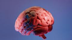
Scientists at the Sudha Gopalakrishnan Brain Centre (SGBC) at the Indian Institute of Technology, Madras (IIT-M) have created what they describe as the world's most detailed three-dimensional atlas of the human brainstem at cellular resolution. The digital map, named Anchor (Atlas of Neurochemical Characterisation of the Human Brainstem with 3D Reconstruction), allows users to seamlessly zoom from MRI scans of the whole brain down to individual neurons.
The atlas combines more than 500 tissue sections from foetal, childhood and adult brains. Built from high-resolution microscope images rather than costlier molecular techniques, it identifies over 200 clusters of brain cells and nerve pathways. Eight chemical markers help distinguish different cell types, providing one of the clearest pictures yet of this vital but poorly understood brain region.
The brainstem, though a small part of the brain, controls breathing, heartbeat, sleep, wakefulness and movement. Its densely packed architecture has long frustrated detailed mapping. Anchor links two largely separate worlds: medical imaging, which shows the whole brain, and cellular pathology, which reveals it one cell at a time.
Rebecca Folkerth, a neuropathologist affiliated with Harvard Medical School and New York University who collaborated on the project, said the atlas fulfills a dream she had early in her career: to have brain scans match the brain's microscopic anatomy. She noted that currently, for Alzheimer's disease, only 15 to 20 sections are typically examined - a fraction of a percent of the whole organ.
The atlas is not a diagnostic tool but a freely available online reference. It could help neurosurgeons navigate the brainstem more safely and improve understanding of disorders like Parkinson's disease, stroke, Alzheimer's disease and sudden infant death syndrome (SIDS).
Project head Mohanasankar Sivaprakasam noted that while animal brains have been mapped in detail, the human brain remains under-charted due to scarce tissue studies. SGBC now plans to image over 100 whole human brains across different life stages and neurological conditions, creating a reference library that could reveal how disease reshapes the brain cell by cell.