Eastern Hospital Uses New Method to Destroy Rare Benign Tumor in Teenager
For the first time, the Radiology Center of Eastern Hospital used high-precision CT guidance to destroy a rare bone tumor in a 13-year-old boy without surgery.
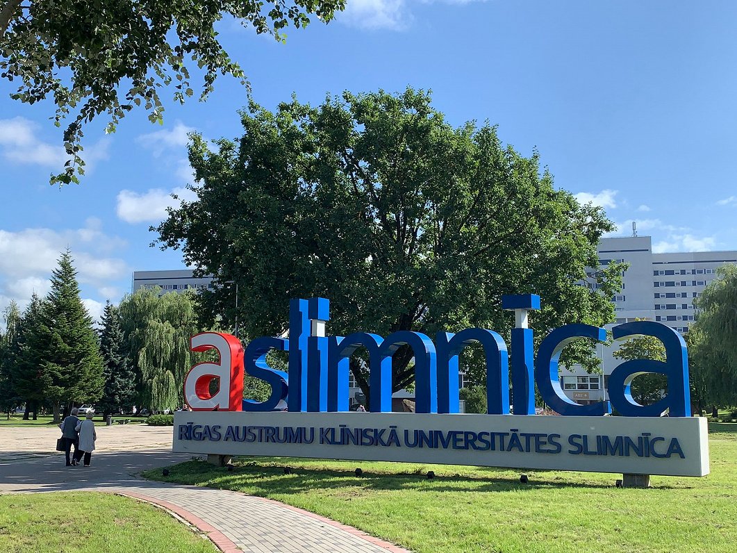
Medical professionals at the Radiology Center of Eastern Hospital have achieved a significant milestone by applying high-precision computed tomography (CT) guidance for the first time to destroy a rare benign bone tumor in an adolescent. This innovative approach allowed the patient—a 13-year-old boy—to avoid extensive surgical intervention. The doctors expressed particular satisfaction because the patient is a teenager, making the less invasive procedure especially valuable. The new method involves accurately locating the tumor using CT imaging and then precisely targeting it for destruction while sparing surrounding healthy tissue. This success opens up new possibilities for treating similar tumors in the future, particularly in children and adolescents, for whom surgery may carry long-term complications. Hospital representatives emphasize that the method is safe and effective, and they plan to use it for other patients with comparable diagnoses. The case marks a significant step forward for Latvian medicine, showcasing the potential of modern technology.
The procedure was completed without complications, and the boy is already recovering. Without the new technique, a major operation would have been necessary, involving a longer rehabilitation period and increased risks. Thanks to the precise CT guidance, the tumor was completely destroyed while preserving bone structure and function. This experience could serve as a model for other medical institutions in Latvia, encouraging the adoption of minimally invasive methods.